
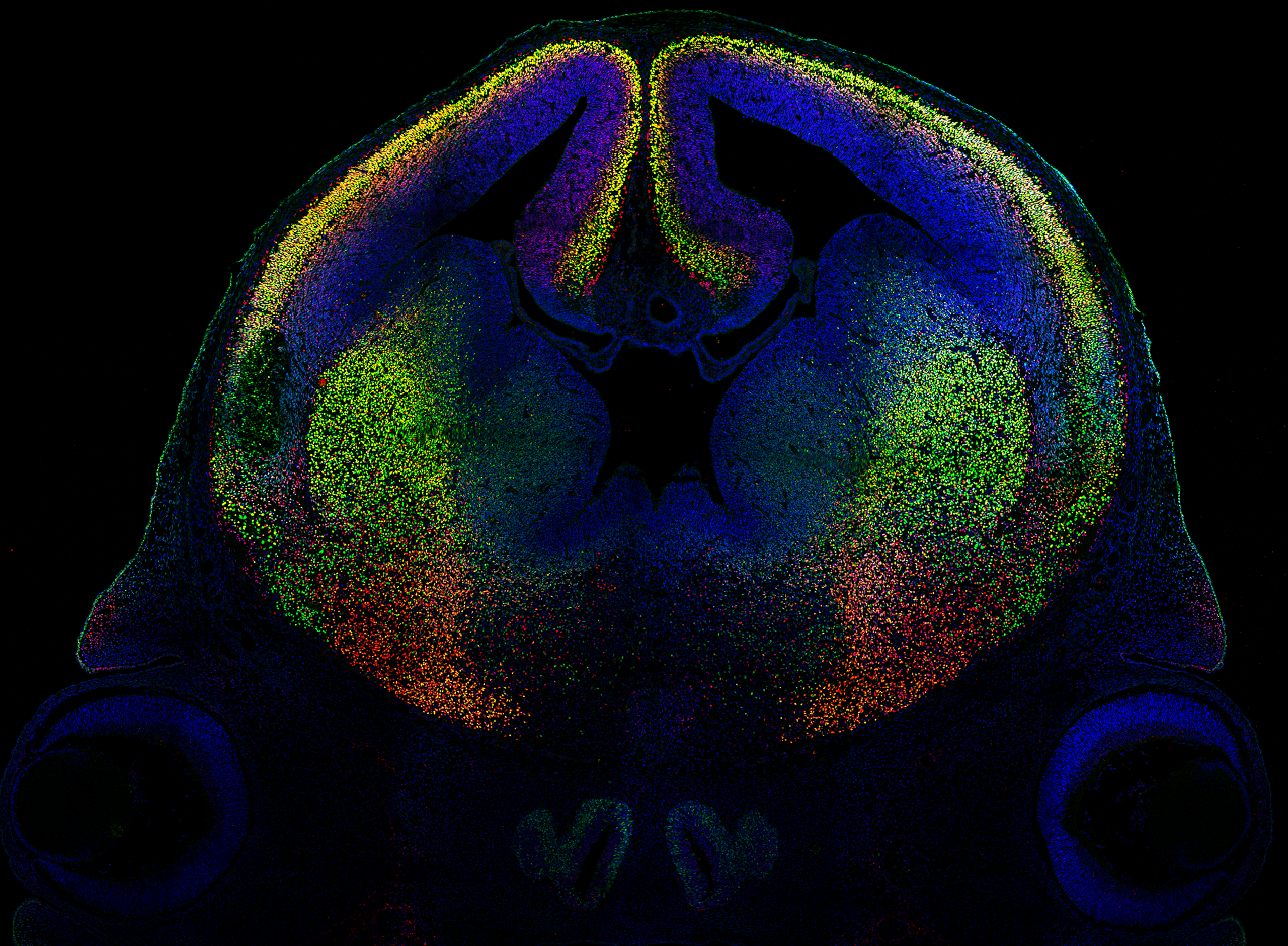
Mouse brain (E14.5), coronal section, Bcl11a (red), Bcl11b (green), DAPI (blue)
Mouse brain (E14.5), coronal section, Bcl11a (red), Bcl11b (green), DAPI (blue)
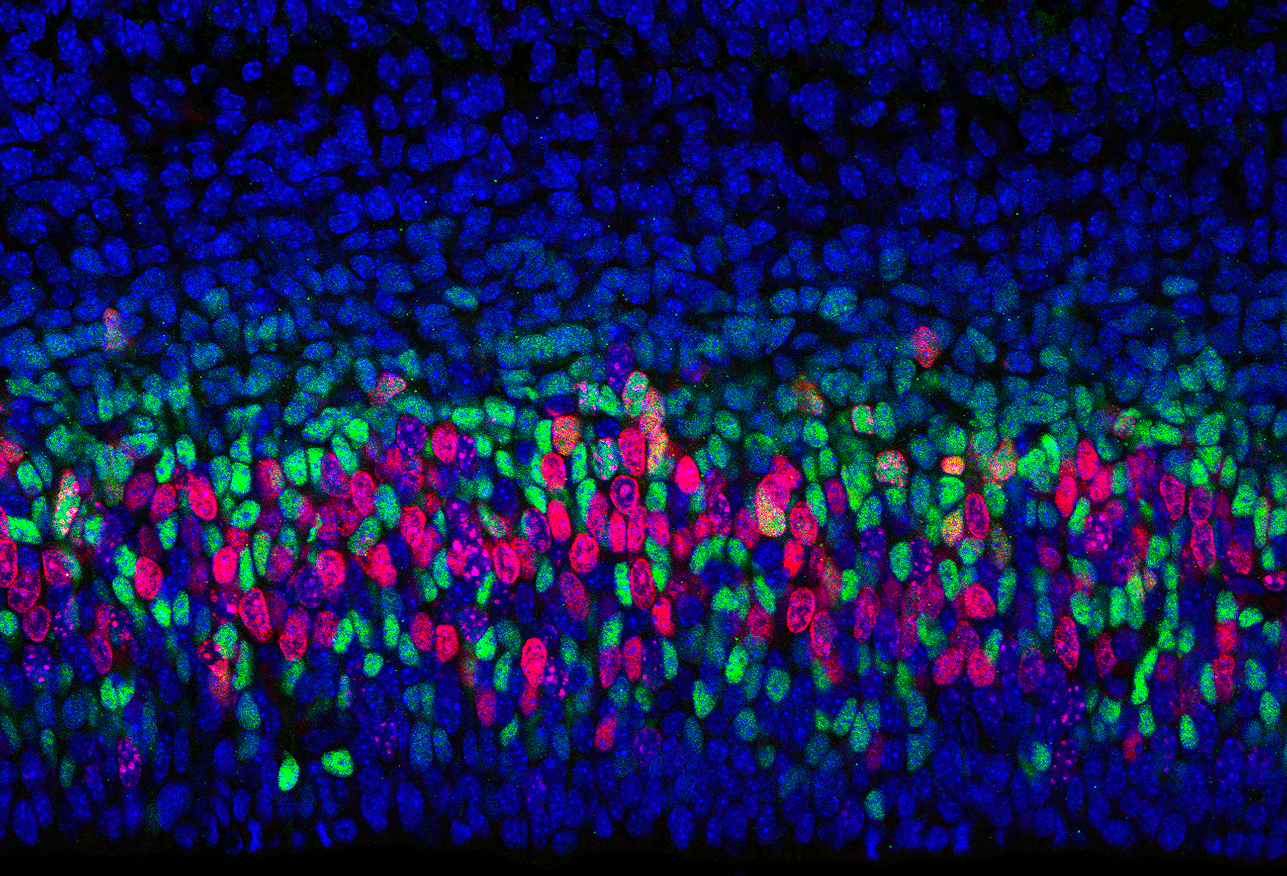
Mouse neocortex (E14.5), EdU (red), Tbr2 (green), DAPI (blue)
Mouse neocortex (E14.5), EdU (red), Tbr2 (green), DAPI (blue)
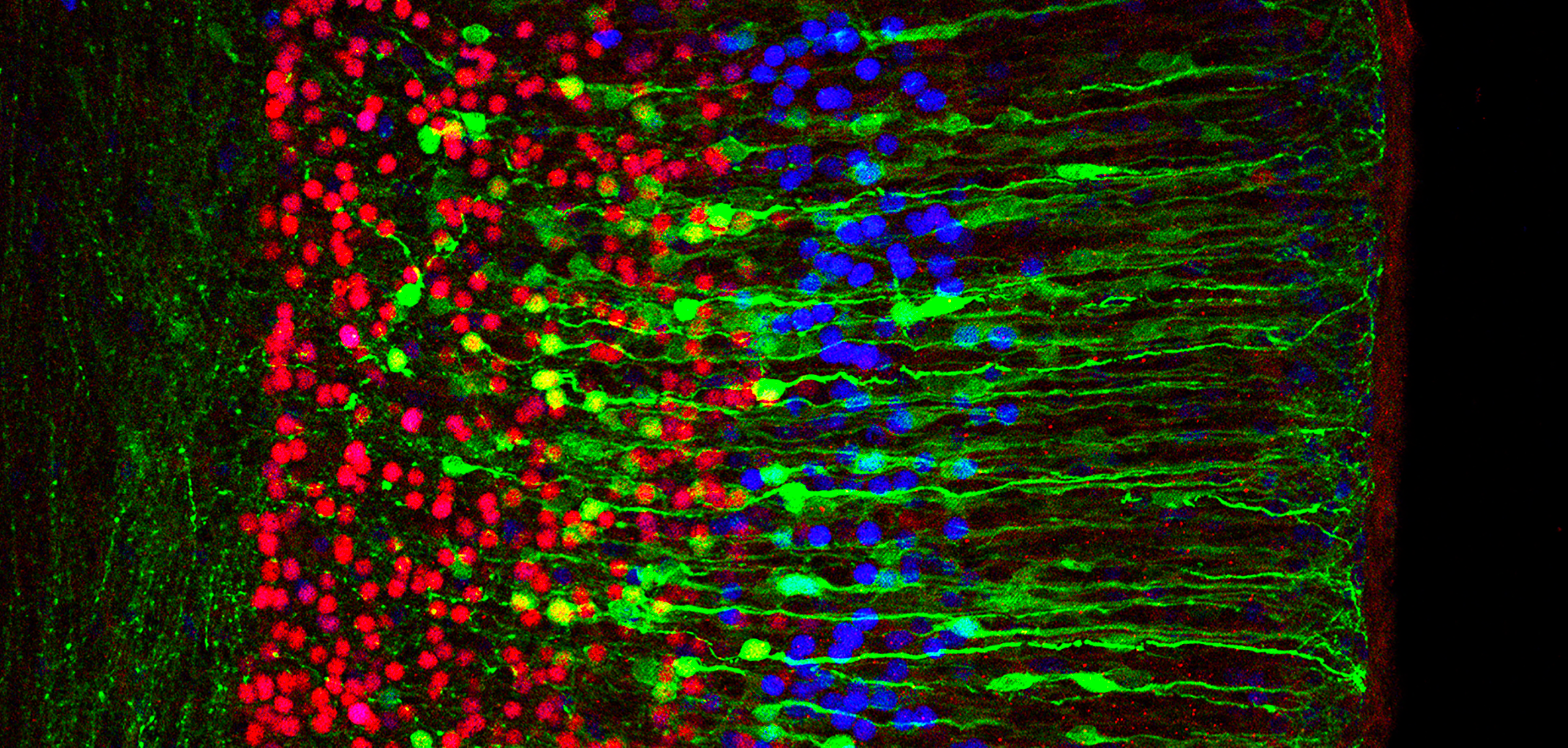
Mouse neocortex, (E18.5), iuE E14.5 -> E18.5: GFP (green), Fog2 (red), Bcl11b (blue)
Mouse neocortex, (E18.5), iuE E14.5 -> E18.5: GFP (green), Fog2 (red), Bcl11b (blue)
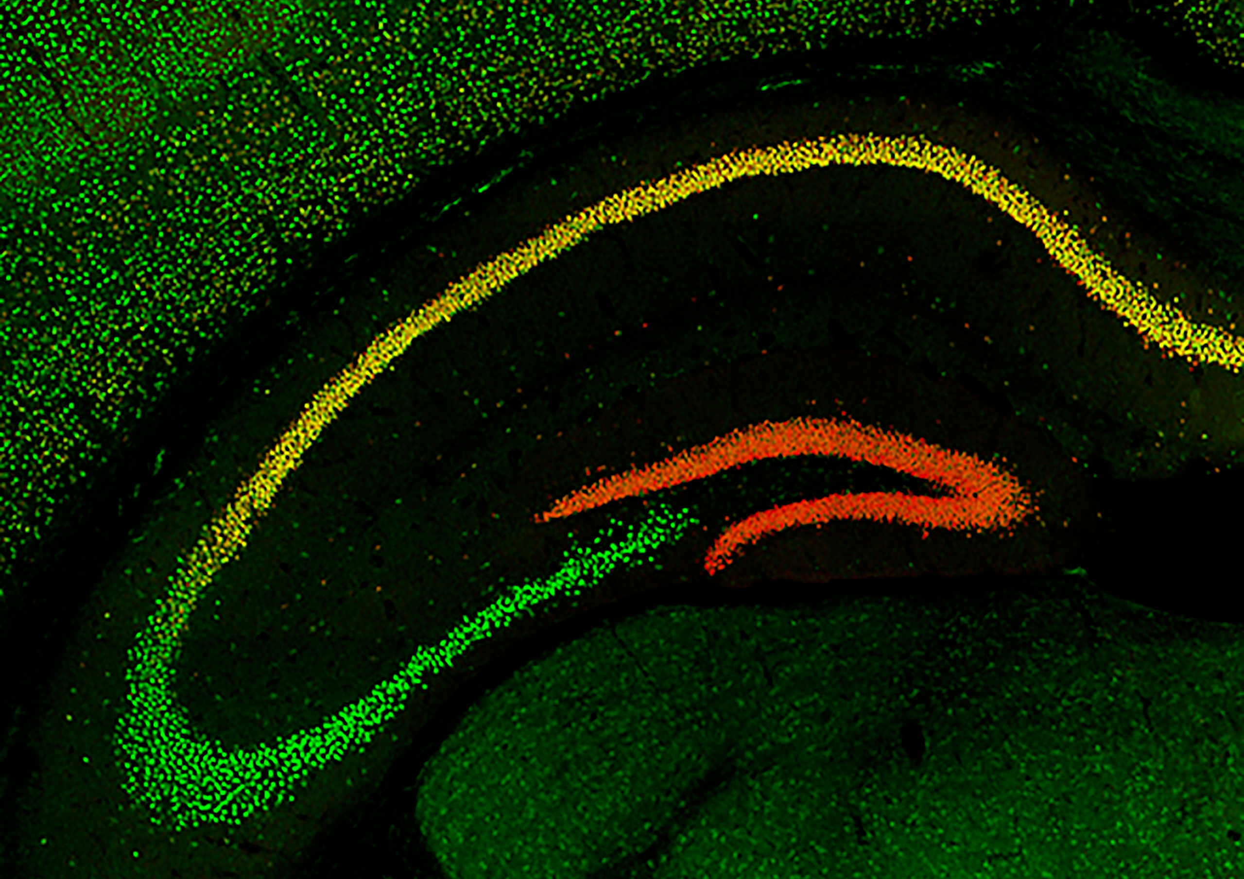
Mouse hippocampus, P30, Bcl11b (red), Bcl11a (green)
Mouse hippocampus, P30, Bcl11b (red), Bcl11a (green)
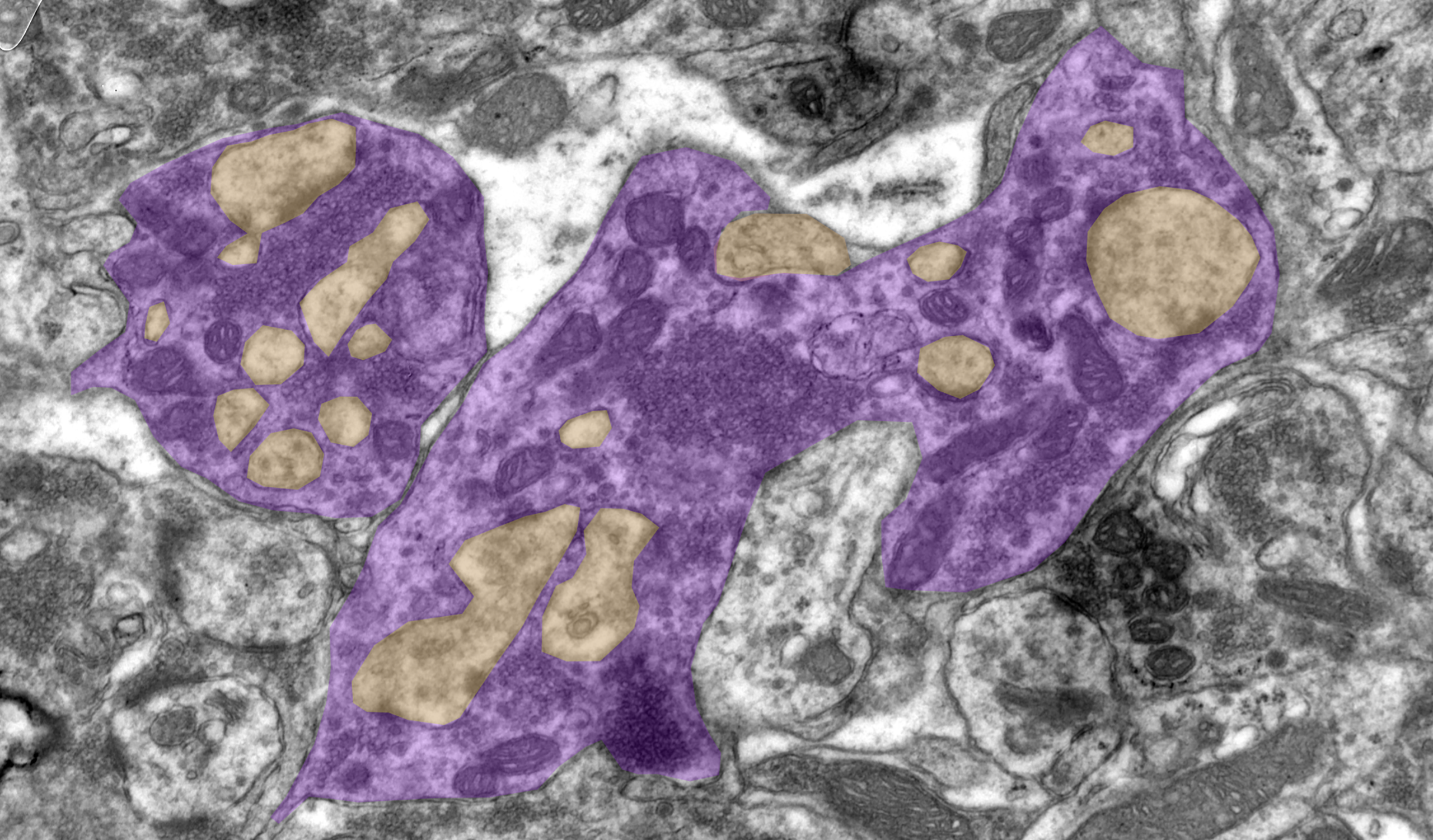
Electron microscopy image of presynaptic dentate gyrus mossy fiber boutons (purple) engulfing postsynaptic CA3 thorny excrescences (yellow) in mouse hippocampus. Image credit: Sigrun Nestel (CC BY 4.0)
Electron microscopy image of presynaptic dentate gyrus mossy fiber boutons (purple) engulfing postsynaptic CA3 thorny excrescences (yellow) in mouse hippocampus. Image credit: Sigrun Nestel (CC BY 4.0)